RESCUE UROLOGY HOSPITAL KENGERI

RECONSTRUCTIVE UROLOGY

Reconstructive Urology

Reconstructive Urology

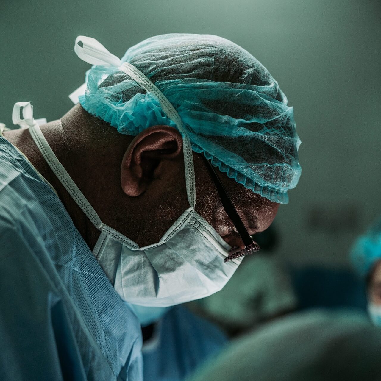
Surgeons specialize in reconstructive urology to treat injuries and conditions affecting the urinary tract and certain reproductive organs. The urology team provides expert care to restore function and help patients to return to their daily activities.
RU is a surgery to restore normal function by repairing, rerouting, or recreating areas of the upper and lower urinary tract and some reproductive organs.

Conditions when Reconstructive Urology is needed
• Traumatic injuries to the urinary tract or reproductive organs
• Birth defects in urinary tract or reproductive organs
• Medical conditions
• Complications from surgery
• Urethral stricture (scar tissue in the urethra that causes narrowing) known as stenosis


SYMPTOMS

  • When there is a problem like a stricture, the bladder has to squeeze harder to eliminate urine.
  • Experience severe pain, excessive strain or prolonged time to empty while urinating.
  • Backup of urine can cause irreversible damage to the bladder and the kidneys.
  • Pelvic fracture or pelvic haematoma.
  • Dark or bloody urine.
  • Prostate infections.
  • Abdominal pain.

Prevalence

  • Babies(0-2) Common
  • Boys (2-18) Common
  • Young adults (19-40) Common
  • Adults (41-60) Common
  • Seniors (60+) Common

Diagnosis and Investigations

Learn about the investigations and the diagnosis

  • A urethral stricture may be detected by an X-ray study or by cystoscopy.
  • The best test is an X-ray study called a retrograde urethrogram.
  • How much scar tissue is present and the length of the stenosis.

Treatment Options

● Dilating the stenosis

● Open surgery (urethroplasty): the affected area of the scar tissue may be removed, and healthy ends of the urethra are then sutured.

● Urethrotomy: involves the use of a scope to cut the stenosis to enlarge the channel. 

Surgery procedure for Urethral Stricture

● Surgery is required to widen or remove the narrowed section of the urethra, if the process of dilation is not responded.
● In urethrotomy, the surgeon uses a special endoscope, a thin instrument with a light embedded at the tip, to make an incision in the urethra for urine to flow freely.
● After the procedure, a catheter is left in place for a few days to divert urine away from urethra thus help the process of healing.
● Pain relief medications are prescribed to help you cope with the pain.
● In urethroplasty, is a surgery done after dilation.
● The surgeon locates and removes the narrowed section of the urethra and joins the two healthy pieces.
● In case the scarred segment is too long to be removed, doctor may use tissue from other parts of the body to recreate the normal size of the urethra .
● In this case also a catheter remains in the urethra for two to three weeks.


Hospice care and stay

● The procedure of urethrotomy does not require an overnight hospital stay, though you may have pain for two weeks after the procedure.
● Urethroplasty is performed in the hospital but as an outpatient procedure. Some people may be discharged the following day, if they need more time for recuperation. 

  • DO YOU HAVE QUESTIONS ABOUT RECONSTRUCTIVE UROLOGY?
  • TELE CONSULT WITH YOUR UROLOGIST

Patient safety is our priority

● Urethral strictures can sometimes return or recur after treatment, usually within the first two years after surgery.
● The longer the stricture, the greater chance of recurrence.
● Doctor continues to monitor urethral functioning by conducting periodic physical examinations and tests as needed.
● After 6 months your doctor may perform a cystoscopy, in which they use a thin scope to see inside the urethra.
● Doctor mar ask for a urethrogram to be conducted, which provides images of the urethra using X-ray or ultrasound. 

  • DO YOU HAVE QUESTIONS ABOUT RECONSTRUCTIVE UROLOGY?
  • TELE CONSULT WITH YOUR UROLOGIST

What's important after discharge?

● Take care to avoid injury to the pelvis by having safe sex.
● Visit your doctor if you have symptoms of urethral stricture.
● Additional dilation procedures or surgery may be needed to treat recurrent urethral stricture. 

  • DO YOU HAVE QUESTIONS ABOUT RECONSTRUCTIVE UROLOGY?
  • TELE CONSULT WITH YOUR UROLOGIST

RECONSTRUCTIVE UROLOGY

Ureteral Injury

Ureteral Injury and Symptoms

Ureteral injury is one of the most serious complications of gynecologic surgery. For these reasons, injuries to the urinary tract, particularly the ureter, are the most common cause for legal action against gynecologic surgeons. These are serious and troublesome and are often associated with significant morbidity.
The formation of uterovaginal fistulas and potential loss of kidney function are the common issues in ureteral injury. They go unnoticed until postoperatively.

Treatment could be surgical and nonsurgical methods.

SYMPTOMS

  • Abdominal pain and swelling
  • Severe flank pain and back pain
  • Blood in urine
  • Drowsiness, decreased alertness including coma
  • Decreased output or inability to urinate
  • Fever
  • Increased heart rate
  • Nausea, vomiting

Prevalence of Ureteral Injury

Incidence

  • Gynecologic procedures account for the majority of ureteral injuries with 64%-82%.
  • Colorectal, vascular pelvic, and urologic surgery account for about 15% - 26%.and 11% - 30% respectively.
  • Cut ureter during Hysterectomy
  • Most of the above cases lead to legal cases against the surgeon.

Diagnosis of Ureteral Injury

Learn about the investigations and the diagnosis

  • The increasing use of laparoscopy as a therapeutic method has increased the risk of ureteral injuries.
  • Complications frequently described include injuries to the large and small bowels, bladder, and blood vessels
  • The diagnosis is made by intravenous pyelography
  • Both intravenous urography and initial urinalysis may be unreliable indicators of ureteral and renal pelvic injury.
  • Satisfactory urinary diversion can be achieved in most cases with an internal ureteral stent.
  • Cystoscopy was used to determine ureteral injuries intraoperatively, post procedure and postoperative periods.
  • Ureteral injury due to blunt and penetrating trauma was diagnosed by retrograde pyelogram which confirmed the disruption of left ureteropelvic junction.

Treamment of Ureteral Injury

● Cystoscopy and retrograde ureteral stent placement or antegrade placement of a ureteral stent
● Laparoscopic techniques for the management of ureteral injuries include Ureteroureterostomy, Ureteroneocystostomy and Boari-flap procedures.
● Clinicians should perform IV contrast enhanced abdominal /pelvic CT with delayed imaging (urogram ) for stable trauma patients with suspected ureteral injuries.
● Clinicians should directly inspect the ureters during laparotomy in patients with suspected ureteral injury who have not had preoperative imaging.
● Surgeon repairs traumatic ureteral lacerations at the time of laparotomy in stable patients
● Surgeons may manage ureteral injuries in unstable patients with a temporary urinary drainage followed by delayed definitive management
● Surgeons should manage traumatic ureteral confusions at the time of laparotomy with ureteral stenting or resection and primary repair depending on ureteral viability and clinical scenario.
● Surgeons should attempt ureteral stent placement in patients with incomplete ureteral injuries diagnosed postoperatively or in a delayed setting.
● Immediate repair can be considered in certain clinical situations if the injury is recognised within one week ( eg, injury located near a surgically closed viscus, such as bowel or vagina, or if the patient is re-explored for other reasons.
● Surgeons should perform percutaneous nephrostomy with delayed repair as needed in patients when stent placement is successful or not possible.
● Surgeons should repair ureteral injuries located proximal to the illiac vessels with primary repair over a ureteral stent, when possible.

Other Surgical Methods Used to Reconstruct the Injured Ureter

● Ureter reimplant
● Psoas hitch surgery
● Boari flap surgery
● Ureter patch or interposition with the appendix
● Ileal ureter
● End-to-end repair
● Robotic urinary tract reconstruction

Hospice care and duration of stay

● After the operation , you may spend some time in the intensive care unit.
● The wound takes 6 weeks before it heals fully.
● The stent helps the ureter to heal, it may be removed after 3 to 4 weeks.

Follow-up after surgery

● You may need to come back after 3 to 6 weeks to get the stent removed which is a quick in office procedure.
● No driving for 2 weeks until the catheter is removed.
● No heavy lifting or strenuous activity for 4 to 6 weeks. 

  • DO YOU HAVE QUESTIONS ABOUT URETERAL INJURY?
  • TELE CONSULT WITH YOUR UROLOGIST

Patient safety is our priority

Risks and complications

● The presence of electrical arcs constitutes an additional risk factor.
● Formation of urinary fistula
● Slight risks of episodes of incontinence( leakage of urine).
● Irritation could be present in and around the area where the stent has been placed.
● Stents can cause discomfort and pain in the bladder, kidneys, groin, urethra and the genitals.
● Some complications of surgery include ureteral obstruction, which can be caused by bleeding or blood clots, bladder spasms, or ureteral ischemia.

  • DO YOU HAVE QUESTIONS ABOUT URETERAL INJURY?
  • TELE CONSULT WITH YOUR UROLOGIST

What's important after discharge?

● Drink 2.5-3 liters of water daily
● Continue taking narcotic pain medicine as needed for moderate to severe pain.
● Take warm baths or heating pad.
● Prevent constipation by regular use of stool softener like Colace, drink juices,eat fibre rich fruits and vegetables.
Meet the Doctor
● When fever more than 101.5    F along with sweats and rigors.
● Bright red blood in urine and large clots.
● Opaque urine that you cannot see through.  

  • DO YOU HAVE QUESTIONS ABOUT URETERAL INJURY?
  • TELE CONSULT WITH YOUR UROLOGIST